IHC 항체 --IgM
양성 대조군:
Tonsil셀룰러 현지화:
Cytoplasm애플리케이션:
IHC-P이차항체:
iVision™카탈로그 번호:
AP0140숙주 종:
Rabbit클론:
polyclonal사양/ml:
1、3、6、0.2(Concentrated)【Product name】
IHC Antibody --IgM
【Packing specification】
| Code |
Clone |
Specifications |
|
AP0140 |
polyclonal |
0.1ml, 0.2ml, 1ml, 1.5ml, 3ml, 6ml, 11ml, 30ml |
【Intended use】
For Research use only. IgM Antibody reagent is intended for use to qualitatively identify IgM by microscopy in sections of FFPE tissue using immunohistochemical detection system.
【Principle】
IgM is one of the predominant surface immunoglobulins on B-lymphocytes. This antibody is useful when differentiating and sub-classifying hematolymphoid neoplasms. Add the primary antibody to bind the antigen on tissue sections, and then use HRP labeled secondary antibody binding primary antibody to form the secondary antibody-primary antibody-antigen complex. When DAB chromogenic solution is added, HRP reacts with enzyme substrate to produce brown insoluble reaction product, which indirectly indicating the existence of antigen.
【Main components】
Immunoglobulin, antibody diluent
【Storage】
Store at 2~8℃ for 18 months。
【Sample requirements】
FFPE tissues are usually cut into sections as thin as 3~5μm with a microtome. These sections are then mounted onto glass slides that are coated with a tissue adhesive.
【Protocol】
1. Sample preparation:Deparaffinize the slides in xylene Ⅰ, Ⅱ, Ⅲ for 5 minutes;Transfer the slides once through 100%, 100%, 95%, 75% alcohols for 2 minutes respectively. Rinse slides with deionized water for 30 seconds.
2. Blocking:Block endogenous peroxidase activity by incubating sections in 3% H2O2 solution at room temperature for 5 minutes to block endogenous peroxidase activity. Rinse the slides with deionized water for 30 seconds.
3. Antigen retrieval:Heat the EDTA Antigen retrieval buffer to 100℃. Then place the slides in the boiled buffer and continue to heat for 15~20 min. Naturally cool down for 30 minutes. Rinse the sample with wash buffer.
4. Primary antibody incubation: Drain the slides. Add primary antibody to tissue, incubate at room temperature for 30 minutes. (use antibody diluent or PBS as control). Wash the slides in PBST for 2 times, 5 minutes for each time. If the Primary antibody is concentrated, please dilute it to RTU(ready to use) according to the information on packing.
5. Secondary antibody: Drain the slides. Add secondary antibody to tissue and incubate at room temperature for 20 minutes. Wash the slides in PBST for 2 times, 5 minutes for each time.
6. DAB:Drain the slides. Add DAB to the tissue and incubate at room temperature for 5 min. Rinse slides with deionized water.
7. Hematoxylin staining:Drain the slides. Add Hematoxylin to the tissue and incubate at room temperature for 5 minutes. Rinse slides with water. Use the acid solution for differentiation. Rinse slides with water.
8. 탈수 : 슬라이드를 75% , 95% , 100% 알코올에 2 분간 탈수 시킨다 . 슬라이드를 말립니다. 마운팅 매체를 사용하여 커버슬립으로 염색된 조직을 덮습니다.
【긍정적인 현지화 】
1. 양성 국소화 : 세포질 .
2. 양성 대조군 : 편도선 .
【주의사항 】
1. 사용하기 전에 지침을 주의 깊게 읽고 키트의 모든 구성 요소에 익숙해지십시오. 작동 중에는 지침을 엄격히 따르십시오.
2. 키트 또는 키트 구성품을 사용한 후에는 사용하지 마십시오.
3. 교육을 받은 전문가만 이 키트를 사용할 수 있습니다. 시약을 취급하는 동안 적절한 실험복과 일회용 장갑을 착용하십시오.
4. 화학물질이 피부, 눈, 점막에 닿지 않도록 하십시오.
5. 입으로 피펫팅하지 마십시오.
6. 사용하지 않은 시약, 사용한 키트 및 폐기물은 현지 규정에 따라 폐기해야 합니다.
【제조사 】_
회사명: Xiamen Talent Biomedical Technology Co., Ltd
주소 : 36100 중국 샤먼시 하이창구 바이오메디컬 파크 웽자오 로드 웨스트 2068호 빌딩 B10 3층 및 4층
전화: +86 592 6315755
이메일 : tlsw@talentbiomedical.com
홈페이지 : www.talentbiomedical.com
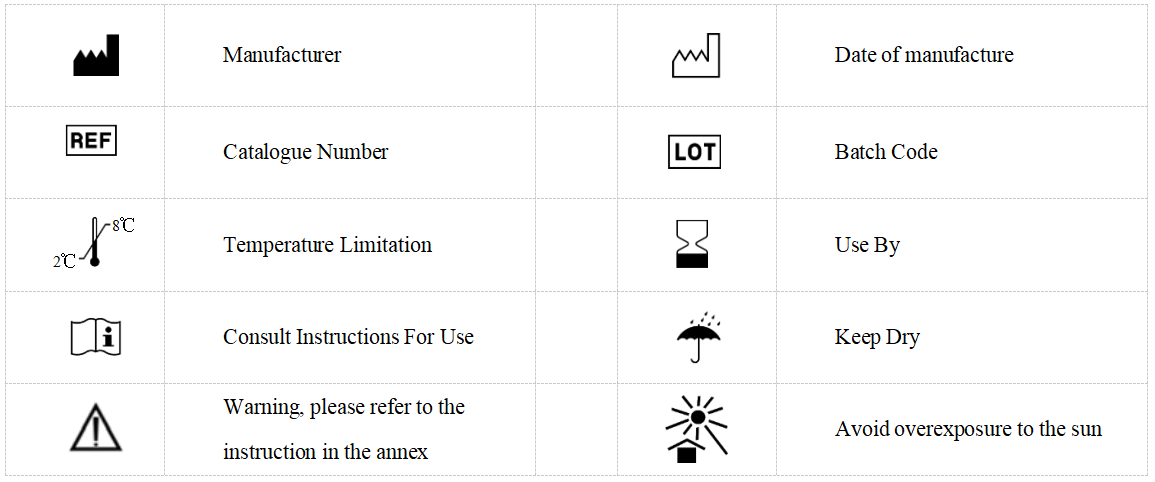
【 기호 】

관련 태그 :